Monday 6 September 2010
Thursday 26 August 2010
In The Shadow Of The Sun

The light of the Sun is the primary source of the free energy that potentiates life on Earth. Solar energy is naturally harnessed by chlorophyll in the leaves of trees and plants; and together with water from the soil and carbon dioxide from the atmosphere, the process of photosynthesis propagates the release of oxygen. As humans, therefore, we have an indirect dependence on the sun and without it, we would all perish. Remarkably, we are also able to harness solar energy by using solar panels, as ways of producing electricity both domestically and commercially.
On Saturday 21st August, as part of the British Film Institute's Film Science season, Dr Adam Rutherford hosted an event called In the Shadow of the Sun at BFI Southbank. Joined by a panel of diverse guests including Film Director/Producer Danny Boyle, particle physicist Professor Brian Cox OBE, curator/artist Honor Harger and solar researcher Dr Lucie Green, the emanating discussions captivated the audience with a personal insight into the making of the 2007 film "Sunshine" and detailed perceptivity into the demeanours of the Sun that most of us are not privileged to know.
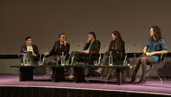 |
| "In the Shadow of the Sun" Panel at BFI Southbank |
A short clip was shown of the opening scenes of "Sunshine", a film set in 2057 about the formidable mission of a team of scientists sent to re-ignite the dying Sun, in order to save Earth. It was the epic scene in which the ships doctor, Searle, is sitting in the observation room, admiring the beauty of the Sun through a filter that is demonstrating only 2% of the Sun's full brightness. He commands the artificial computer, named Icarus, to decrease the filters in order to view the Sun at 4%, but Icarus warns him that viewing the Sun at this intensity would cause permanent damage to the retina, however, it is safe to view at 3.1% but only for thirty seconds. Searle places his sunglasses, the filters are decreased to the absolute safe level and the brightness overpowers him and all those watching the film.
 |
| The observation room, on board Icarus II |
Danny knew he needed a sagacious mind to assist him with these matters. Step up: Professor Brian Cox. This arrangement took a disappointing turn for one of Prof Cox's colleagues after taking the original call from Danny, inviting him to join them on the set as scientific consultant. He must have been excited about this offer since he was approaching retirement, but he soon realised Danny was in fact after Prof Cox when he mentioned that due to his young age he could connect more with the young cast members. Poor old boy.
 |
| Searle preparing to observe the Sun at 3.1% |
 |
| Cillian Murphy as physicist, Robert Capa |
An eminent point made by Prof Cox was one that he re-ignited whilst working on the film, as it reminded him that the fundamental interest that most scientists have in science, were born from an early emotional connection, like the first time a child looks at the moon and stars and realises they want to be an astronaut. Beautiful and very true.
 |
| The Weather Project, at Tate Modern. |
Together with another Sun enthusiast and collectively known as "r a d i o q u a l i a", Honor created a radio station devoted entirely to streaming live radiowaves from the Sun and cosmos, a project called "Radio Astronomy". Sadly, the live feeds no longer run (last live feeds were in 2008), however, the recordings can be retrieved from their archives. A sample clip of what the Sun sounds like was played; fundamentally a series of scratches, hisses and clicks, but awe-inspiring all the same.
Honor also showed us an amazing video by Semiconductor called "Black Rain" which was created using images from the twin satellite, solar mission, STEREO as it scanned the interplanetary space for solar wind and coronal mass ejections heading towards Earth. Here's the full clip, some of which was featured in Prof Cox's Wonders of the Solar System:
Black Rain from Semiconductor on Vimeo.
 |
| "EarthStar" by Haines & Hinterding |
Each lay person could say they knew at least one fact about the Sun; it's a star, it has a massive gravitational pull and that's why our solar system orbits around it, it gives us a tan, it's Ultra Violet (UV) rays damage our skin and could cause skin cancer, it's a big ball of hydrogen and helium gas - that sort of thing. However, other than such superficial facts, not many of us know what is really going on up there, in fact the topic is so arcane, even scientists like Dr Lucie Green who study it everyday are still learning from it. The latest update on the Sun even made the news (here) just this week.
 |
| The chromosphere viewed through UV light |
It is this and the outermost layer, the corona, that Dr Green showed particular interest in, in her talk. Irrationally and sporadically, the chromosphere will project bursts of glowing gases many miles up into the corona layer; sometimes these "prominences" are suspended there for a puzzling amount of time. Other times, these amazing "coronal mass ejections" reach Earth, causing a geomagnetic storm that affects the Earth's magnetosphere by compressing it on the day side and stretching it on the night side. When the night side reinstates it's usual form it, effectively, bounces back an immense protective response to release the solar energy away from Earth. This bouncing back of energy can sometimes be witnessed in the night skies, a beautifully breath-taking display known as the "aurora borealis", or "Northern Lights". Below is a movie clip demonstrating, with the Sun concealed to aid clear viewing, the amazing display of these solar flares blasting their way through the interplanetary space.
These coronal mass ejections and solar flares are extremely powerful and emit masses of radiation. This creates particular problems for satellites and people working in space; as finding yourself in the path of a solar flare would result in radiation sickness and, ultimately, death. In light of this, Dr Green spoke of the up coming problems the latest mission (I missed the name of it - sorry!!), that will be the closest to the Sun ever attempted, so you can imagine what measures are having to be taken to withstand these flares.
As a biomedical scientist, the Sun does not lie within my mainstream of interest so I was amazed at how captivating it became, I particularly believe this is because the panelists represented a multifaceted presentation of various appreciations of the same subject. Now, I most definitely have a deeper interest in this field and will make active attempts to read about the latest research. What an amazing all-rounder of an event, an amazing topic, a great host with truly engaging guests.
Additional reference points other that the in-text links provided:
http://www.flickr.com/photos/britishfilminstitute/4919764966/in/photostream/
http://www.physics.ucla.edu/pdg/Homepage/equinoxprom_eit.jpg
http://www.dlr.de/en/DesktopDefault.aspx/tabid-5170/8702_read-17935/8702_page-3/gallery-1/gallery_read-Image.1.9670/
http://www.solarviews.com/eng/sun.htm
http://www.dlr.de/en/DesktopDefault.aspx/tabid-5170/8702_read-17935/8702_page-3/gallery-1/gallery_read-Image.1.9670/
http://www.solarviews.com/eng/sun.htm
Tuesday 24 August 2010
Oncogenesis: Moving up and down a NOTCH.
Oncogenesis is defined as the progression of cytological, genetic and cellular changes that ultimately lead to uncontrolled proliferation and malignant transformation. In normal tissue, regulatory processes recognise when cells must undergo proliferation, differentiation, repair or death to eliminate renegade cells and prevent the threat of transformation. Factors that disrupt this regulation contribute to oncogenesis but more than one “hit” is required to actually cause cancer.
The Notch signalling pathway (see below) is a direct paracrine, cell-to-cell signalling pathway that controls apoptosis, cell cycle, cell proliferation, cell differentiation, neurogenesis and transcription and due to these important regulatory roles, aberrant Notch signalling is considered to assist in cancer progression. This blog looks at how one aspect of the Notch signalling pathway, the Notch1 receptor, may have an important role in oncogenesis, considering both oncogenic and tumour suppressive perspectives.
The Notch receptor is an evolutionary conserved transmembrane glycoprotein with an amino-terminal extracellular domain (ECD) and a carboxyl-terminal intracellular domain (ICD). It is initially synthesised as an inactive precursor which is cleaved by Furin protease in the Golgi apparatus (first cleave known as S1) to produce the Notch ICD (NICD) and Notch ECD (NECD). These domains then become O-fucosylated by O-fucosyl transferase and glycosylated by Fringe before becoming integrated into the cell membrane. The transmembrane Notch receptor becomes activated upon the binding of transmembrane ligands from the Delta and Serrate/Jagged (DSL) families. E3 ligases (Neuralised and Mindbomb) facilitate epsin-dependent endocytosis of the bound ligand to expose the second Notch cleavage site (S2) to metalloproteases ADAM (a disintegrin and metalloptoreinase) 10 & 17 to release NECD. Cleavage of NECD, in turn, triggers the final cleavage (S3) within the cell membrane by the g-secretase complex (Presenilin-Nicastrin-APH1-PEN2) and releases NICD. NICD then translocates to the nucleus where it assembles with an ubiquitous DNA-binding protein of the CBF1/RBP-Jk, Su(H) and Lag-1 (CSL) family and other co-activators such as Mastermind-like proteins (MAML) to become a transcription complex which target genes (see table) that ultimately decide the fate of the cell.
Humans have four Notch receptors, Notch1, Notch2, Notch3 and Notch4 and five equally conserved canonical Notch DSL ligands and these are Jagged1, Jagged2 and Delta-like 1 (Dll1), Delta-like 3 (Dll3) and Delta-like 4 (Dll4). Each Notch receptor and DSL ligand combination determines a particular Notch-mediated response; their regulation is extremely important for preventing abnormal cells from transformation and so, as with any biomolecular pathway, aberrant Notch signalling can have extremely compromising oncogenic outcomes but as the following explains, Notch can also result in tumour suppression.
Oncogenic roles of Notch signalling
In the haematopoietic system, Notch1 activation is important for T-cell lineage specification and development, demonstrated by Notch1 deletion causing failure of T-cells to develop and increased expression of Notch1 resulting in double positive T-cell’s and inhibition of B-cell development in the bone marrow. In one form of T-cell acute lymphoblastic leukaemia (T-ALL), a translocation t(7;9)(q34;q34.3) juxtaposes a truncated carboxyl terminal of Notch1 to the T-cell receptor-b (TCRb) promoter/enhancer gene (right). Subsequent expression of this truncated Notch1, transcribes only for the NICD portion, resulting in a mutant form lacking the heterodimerisation domain and Lin12-Notch repeats (HD-LNR) that function to prevent spontaneous activation in the absence of a ligand-receptor interaction. The truncated Notch1 protein varies depending on the exact translocation and translation initiation sites; as some remain as transmembrane proteins that require g-secretase processing for activation, whereas others are freely activated within the cytosol. The role of Notch signalling due to the translocation and subsequent over-expression of the NICD component is oncogenic, evidenced by ectopic T-cells developing into aggressive, proliferating monoclonal T-cell tumours expressing N1ICD.
Approximately 40% of human T-ALLs contain mutations within exons 26 and 27, which encode for the HD region and this demonstrates its importance in stabilising Notch signalling. Further Notch1 mutations, other than within the HD region, have been identified in T-ALL, including the PEST region, PEST and HD combined, juxtamembrane and transactivation domains, each with a consequent oncogenic outcome. Additional evidence for an oncogenic role of Notch1 in T-ALL is demonstrated by over-expression of c-Myc (only) proving insufficient in causing T-ALL, yet an additional Notch1 mutation (induced by viral insertions) targeting c-Myc subsequently induced tumour progression.
Mutations, however, are not the sole means for Notch1 contribution to oncogenesis. Wild type Notch1 signalling may be a downstream effector of oncogenic Ras as increased Notch1 signalling, simply through Ras up-regulating the Notch ligand Delta-1, provided a complimentary environment for sustaining mammary tumours. Another non-mutation-derived oncogenicin which an increased activity of the pathway through increased expression of Notch ligands and receptors, was induced by a hypoxic environment. Hypoxia is a shared attribute of solid tumours, with many over-expressing Hypoxia inducible factor 1a (HIF-1a), known to initiate further invasion, angiogenesis and distant metastasis. The research suggested the effect of hypoxia on Notch signalling was due to the accumulation of HIFs that synergistically activated transcription of both HES1 and HEY1 with the Notch coactivator MAML1 and that Notch activation correlated with hypoxia-mediated breast cancer cell invasion and metastasis. A similar study of adenocarcinomas of the lung showed that, under hypoxia, inhibition of Notch1 signalling induces apoptosis of the tumour cells and that Notch1 activation maintains an oncogenic role by activating Akt-1 through repression of phosphatase and tensin (PTEN) homologue and induction of insulin-like growth factor-1 receptor (IGF-1R) where IGF-1R functions to protect the tumour cells from g-secretase inhibitor (GSI) toxicity, which would normally induce apoptosis. Earlier research also compliments these findings as they found a dependency of malignant mesothelioma cells, under hypoxia, for increased Notch1 activity to increase the prosurvival phosphatidylinositol 3-kinase/Akt/mammalian target of rapamycin (PI3K/Akt/mTOR) signalling pathway and maintain their tumourigenic state.
Notch1 activation resulted in a significant in vitro and in vivo growth inhibition of hepatocellular carcinoma (HCC) cell line SMMC7721, through decreased expression of cyclin A1, cyclin D1, cyclin E, CDK2, decreased phosphorylation of Rb and increased expression of p21 (cell cycle arrest) together with p53 up-regulation and Bcl-2 down-regulation (apoptosis) (see below).
Notch1 is also expressed in low levels in HCC and this correlated with high levels of beta-catenin, suggesting that up-regulation of Notch1 signalling in this context maintains tumour suppression. Comparatively, however, some studies concluded that Notch1 in fact has an oncogenic role in HCC with HCC cells, when compared to normal adjacent hepatocytes, showing an up-regulation of cytoplasmic Notch1; sometimes together with increased expression of HES-1 gene also. The oncogenic role of Notch1 in HCC was demonstrated by exposing the tumour cells to a Notch1 inhibitor (curcumin), which resulted in a dose-dependent N1ICD reduction, HCC cell growth inhibition and apoptosis, including a 40% reduction in tumour growth in vivo. Presently, the determining factors that dictate which role Notch1 undertakes in HCC is still uncertain.
In cases of squamous metaplasia of cervical columnar epithelium in early Human Papilloma Virus (HPV) induced cervical intraepithelial neoplasia (CIN) I-III or well differentiated superficial cervical carcinomas, Notch1 expression is increased, leading to suppression of c-Fos protein expression and induction of Fra-1. The result is a negative regulation of HPV dependent transcription, activation of p53 and subsequent p21 activation that, in turn, initiates differentiation and repression of both Wnt and Shh signalling pathways that ultimately lead to tumour suppression. In low grade CIN, HPV-E6 and HPV-E7 genes demonstrate low expression, however, in high-grade malignancies, expression is high and to maintain the transformed malignant phenotype, this is sustained by down-regulating Notch1.
The oncogenic activity of HPV-E6 suppresses p53 by targeting it for ubiquitination and degradation whereas HPV-E7 inhibits p21 and functionally inactivates p105-Rb, thus providing the virus with complimentary alterations that assist cell transformation and tumour progression (above). Another oncogenic role of Notch1 in cervical cancers is playing part to two pro-oncogenic effector mechanisms, namely PI3K/Akt pathway activation and up-regulation of c-Myc that correlate with the dose-dependent effects of Notch1 on papillomavirus oncogenes.
Opposing the tumour suppressive crosstalk of Notch and Wnt signalling pathways seen in early HPV lesions, is the oncogenic role in human intestinal tumours, where the majority show loss of the adenomatous polyposis coli (APC) gene, a key negative regulator of Wnt pathway. Initial research into murine adenomas showed that Notch activation in mice heterozygous for loss-of-function APC gene resulted in a significant increase in number of adenomas compared to the heterozygote littermates lacking Notch activation. The crosstalk between Notch and Wnt signalling was evidenced by the majority of adenoma cells expressing active proliferation together with activated Notch transgene and the nuclear localisation of b-catenin. When immunohistochemistry demonstrated that human adenomas expressed higher levels of Notch1 and Notch targets Hes1 and Hey1 than adenocarcinomas, it was concluded that increased Notch signalling may contribute to the transformation of benign adenomas to colorectal cancer.
In summary, from the research studied in this essay, Notch1 has shown oncogenic roles through over-expression of NICD (T-ALL), increased wild–type activity (breast cancer) and a down-regulation of Notch1 (high-grade cervical cancer). In these cases, Notch1 alone may not be the sole cause of oncogenesis, but rather a contributor along with other oncogenic influences and, evidently, oncogenic Notch1 is not necessarily a mutant gene or protein, as simple up-regulation or over stimulation from upregulated ligands can be responsible in wild-type cases. Tumour suppressive roles of Notch1 activation have also been demonstrated with its absence or dysregulation resulting in hepatocellular carcinoma, BCCs and BCC-like lesions, although contradicting ideas exist with regards to HCC, possibly indicating an unknown cross talk with other signalling pathways or some undetermined link to specific HCC cancer cells. Notch activity outcome is also shown to be context-specific such as the increased activity seen in breast cancer cell invasion and metastasis in response to increased HIFs, as well as the varied stages of HPV-induced lesions in cervical cancer. An important aspect of the role of Notch is how interactions with other signal transducing pathways contribute to oncogenesis. Again, there is no strict rule for Notch1 activity outcome with a particular pathway, as shown with Wnt cross talk, where in early HPV lesions, an increase in Notch1 activity results in tumour suppression, but in human intestinal tumours this is oncogenic. Similarly, Notch1 crosstalk with the p53 pathway also showed varied outcomes, as increased Notch1 activity resulted in decreased p53 in T-ALL and mouse embryonic fibroblast tumours and decreased Notch1 activity resulted in decreased p53 activity in late HPV lesions.
Weijzen S, Rizzo P, Braid M, Vaishnav R, Jonkheer SM, Zlobin A, Osborne BA, Gottipati S, Aster JC, Hahn WC, Rudolf M, Siziopikou K, Kast WM, Miele L (2002) Activation of Notch-1 signaling maintains the neoplastic phenotype in human Ras-transformed cells. Nat Med 8: 979 – 986.
The Notch signalling pathway (see below) is a direct paracrine, cell-to-cell signalling pathway that controls apoptosis, cell cycle, cell proliferation, cell differentiation, neurogenesis and transcription and due to these important regulatory roles, aberrant Notch signalling is considered to assist in cancer progression. This blog looks at how one aspect of the Notch signalling pathway, the Notch1 receptor, may have an important role in oncogenesis, considering both oncogenic and tumour suppressive perspectives.
 |
| The Notch signalling pathway, showing cleavage sites, S1-S3 (red underline), ligand activation and subsequent translocation of the Notch Intracellular Domain (NICD) to the nucleus where it becomes part of a transcription activation complex. A closer look at the Notch1 receptor shows the various domains in detail (see abbreviations for further details). |
The Notch receptor is an evolutionary conserved transmembrane glycoprotein with an amino-terminal extracellular domain (ECD) and a carboxyl-terminal intracellular domain (ICD). It is initially synthesised as an inactive precursor which is cleaved by Furin protease in the Golgi apparatus (first cleave known as S1) to produce the Notch ICD (NICD) and Notch ECD (NECD). These domains then become O-fucosylated by O-fucosyl transferase and glycosylated by Fringe before becoming integrated into the cell membrane. The transmembrane Notch receptor becomes activated upon the binding of transmembrane ligands from the Delta and Serrate/Jagged (DSL) families. E3 ligases (Neuralised and Mindbomb) facilitate epsin-dependent endocytosis of the bound ligand to expose the second Notch cleavage site (S2) to metalloproteases ADAM (a disintegrin and metalloptoreinase) 10 & 17 to release NECD. Cleavage of NECD, in turn, triggers the final cleavage (S3) within the cell membrane by the g-secretase complex (Presenilin-Nicastrin-APH1-PEN2) and releases NICD. NICD then translocates to the nucleus where it assembles with an ubiquitous DNA-binding protein of the CBF1/RBP-Jk, Su(H) and Lag-1 (CSL) family and other co-activators such as Mastermind-like proteins (MAML) to become a transcription complex which target genes (see table) that ultimately decide the fate of the cell.
 |
| NICD target genes for transcription and their role |
Humans have four Notch receptors, Notch1, Notch2, Notch3 and Notch4 and five equally conserved canonical Notch DSL ligands and these are Jagged1, Jagged2 and Delta-like 1 (Dll1), Delta-like 3 (Dll3) and Delta-like 4 (Dll4). Each Notch receptor and DSL ligand combination determines a particular Notch-mediated response; their regulation is extremely important for preventing abnormal cells from transformation and so, as with any biomolecular pathway, aberrant Notch signalling can have extremely compromising oncogenic outcomes but as the following explains, Notch can also result in tumour suppression.
Oncogenic roles of Notch signalling
 |
| An illustration of the translocation of the truncated Notch1 in T-ALL |
Approximately 40% of human T-ALLs contain mutations within exons 26 and 27, which encode for the HD region and this demonstrates its importance in stabilising Notch signalling. Further Notch1 mutations, other than within the HD region, have been identified in T-ALL, including the PEST region, PEST and HD combined, juxtamembrane and transactivation domains, each with a consequent oncogenic outcome. Additional evidence for an oncogenic role of Notch1 in T-ALL is demonstrated by over-expression of c-Myc (only) proving insufficient in causing T-ALL, yet an additional Notch1 mutation (induced by viral insertions) targeting c-Myc subsequently induced tumour progression.
Mutations, however, are not the sole means for Notch1 contribution to oncogenesis. Wild type Notch1 signalling may be a downstream effector of oncogenic Ras as increased Notch1 signalling, simply through Ras up-regulating the Notch ligand Delta-1, provided a complimentary environment for sustaining mammary tumours. Another non-mutation-derived oncogenicin which an increased activity of the pathway through increased expression of Notch ligands and receptors, was induced by a hypoxic environment. Hypoxia is a shared attribute of solid tumours, with many over-expressing Hypoxia inducible factor 1a (HIF-1a), known to initiate further invasion, angiogenesis and distant metastasis. The research suggested the effect of hypoxia on Notch signalling was due to the accumulation of HIFs that synergistically activated transcription of both HES1 and HEY1 with the Notch coactivator MAML1 and that Notch activation correlated with hypoxia-mediated breast cancer cell invasion and metastasis. A similar study of adenocarcinomas of the lung showed that, under hypoxia, inhibition of Notch1 signalling induces apoptosis of the tumour cells and that Notch1 activation maintains an oncogenic role by activating Akt-1 through repression of phosphatase and tensin (PTEN) homologue and induction of insulin-like growth factor-1 receptor (IGF-1R) where IGF-1R functions to protect the tumour cells from g-secretase inhibitor (GSI) toxicity, which would normally induce apoptosis. Earlier research also compliments these findings as they found a dependency of malignant mesothelioma cells, under hypoxia, for increased Notch1 activity to increase the prosurvival phosphatidylinositol 3-kinase/Akt/mammalian target of rapamycin (PI3K/Akt/mTOR) signalling pathway and maintain their tumourigenic state.
Tumour suppressive role of Notch signalling
NOTCH1 plays an important tumour suppressive function in the context of epithelial tumours, demonstrated widely using mouse skin models. Loss of Notch1 has the same outcome as loss of p21WAF/Cip1 (cell cycle inhibitor at S phase) on both stem cell populations of keratinocytes and ras- and chemically-induced carcinogenesis of the skin. In the basal cells of the skin Notch1 signalling halts the cell cycle via increasing p21WAF/Cip1 expression, which promotes terminal differentiation and Notch1-/- mice demonstrated spontaneous skin lesions similar to basal cell carcinomas (BCCs). Interestingly, BCCs in humans, which are commonly linked to aberrant Sonic-Hedgehog (Shh) signalling, showed reduced Notch1 expression associated with increased and sustained Gli2 expression, which is a downstream target of the Shh signalling pathway. A tumour suppressive role for Notch1 has been demonstrated in mouse skin as Notch1 ablation resulted in epidermal hyperplasia followed by skin tumours and facilitated chemical-induced skin carcinogenesis, also by means of sustained Gli2 expression. Also identified is inactivation of Notch1 reduced the beta-catenin/Wnt (Wingless/Integration) signalling pathway, thus preventing cells from undergoing differentiation.Notch1 activation resulted in a significant in vitro and in vivo growth inhibition of hepatocellular carcinoma (HCC) cell line SMMC7721, through decreased expression of cyclin A1, cyclin D1, cyclin E, CDK2, decreased phosphorylation of Rb and increased expression of p21 (cell cycle arrest) together with p53 up-regulation and Bcl-2 down-regulation (apoptosis) (see below).
 |
| The tumour suppressive effect of increased Notch1 activity on cell cycle and apoptosis in Human Hepatocellular Carcinoma, demonstrating a G0/G1 cell cycle arrest and increased apoptosis, concluded as Notch1 activation induced. |
Notch1 is also expressed in low levels in HCC and this correlated with high levels of beta-catenin, suggesting that up-regulation of Notch1 signalling in this context maintains tumour suppression. Comparatively, however, some studies concluded that Notch1 in fact has an oncogenic role in HCC with HCC cells, when compared to normal adjacent hepatocytes, showing an up-regulation of cytoplasmic Notch1; sometimes together with increased expression of HES-1 gene also. The oncogenic role of Notch1 in HCC was demonstrated by exposing the tumour cells to a Notch1 inhibitor (curcumin), which resulted in a dose-dependent N1ICD reduction, HCC cell growth inhibition and apoptosis, including a 40% reduction in tumour growth in vivo. Presently, the determining factors that dictate which role Notch1 undertakes in HCC is still uncertain.
Varied oncogenic and tumour suppressive roles of Notch Signalling
In cervical cancer, Notch1 signalling is known to have both oncogenic and tumour suppressive qualities.  |
| "Double edged sword" roles of Notch1 in cervical cancer |
The oncogenic activity of HPV-E6 suppresses p53 by targeting it for ubiquitination and degradation whereas HPV-E7 inhibits p21 and functionally inactivates p105-Rb, thus providing the virus with complimentary alterations that assist cell transformation and tumour progression (above). Another oncogenic role of Notch1 in cervical cancers is playing part to two pro-oncogenic effector mechanisms, namely PI3K/Akt pathway activation and up-regulation of c-Myc that correlate with the dose-dependent effects of Notch1 on papillomavirus oncogenes.
Cross talk between signalling pathways.
Notch signalling also plays a role in oncogenesis by means of cross talking with other signal transducing pathways. The example above shows how increased Notch1 activity in early HPV-induced lesions repress both Wnt and Shh pathways and how late-stage cervical cancer leads to ubiquitination of p53 as a result of down-regulated Notch1 signalling, so can be considered as an indirect control of these pathways. Another indirect method of p53 suppression is shown in a form of T-ALL, in which up-regulated Notch1 activity decreases the activity of p14(ARF) which is a negative regulator of MDM2 (an E3 ubiquitin ligase), subsequently leading to increased activity of MDM2 which targets p53 for degradation. A third example shows how deletion of FBXW7/SEL-10 (F-box protein Fbxw7 that mediates the ubiquitination of cell cycle regulator proteins), increases Notch1 activity in mouse embryonic fibroblasts, causing tumour growth suppression by up-regulation of p53.Opposing the tumour suppressive crosstalk of Notch and Wnt signalling pathways seen in early HPV lesions, is the oncogenic role in human intestinal tumours, where the majority show loss of the adenomatous polyposis coli (APC) gene, a key negative regulator of Wnt pathway. Initial research into murine adenomas showed that Notch activation in mice heterozygous for loss-of-function APC gene resulted in a significant increase in number of adenomas compared to the heterozygote littermates lacking Notch activation. The crosstalk between Notch and Wnt signalling was evidenced by the majority of adenoma cells expressing active proliferation together with activated Notch transgene and the nuclear localisation of b-catenin. When immunohistochemistry demonstrated that human adenomas expressed higher levels of Notch1 and Notch targets Hes1 and Hey1 than adenocarcinomas, it was concluded that increased Notch signalling may contribute to the transformation of benign adenomas to colorectal cancer.
In summary, from the research studied in this essay, Notch1 has shown oncogenic roles through over-expression of NICD (T-ALL), increased wild–type activity (breast cancer) and a down-regulation of Notch1 (high-grade cervical cancer). In these cases, Notch1 alone may not be the sole cause of oncogenesis, but rather a contributor along with other oncogenic influences and, evidently, oncogenic Notch1 is not necessarily a mutant gene or protein, as simple up-regulation or over stimulation from upregulated ligands can be responsible in wild-type cases. Tumour suppressive roles of Notch1 activation have also been demonstrated with its absence or dysregulation resulting in hepatocellular carcinoma, BCCs and BCC-like lesions, although contradicting ideas exist with regards to HCC, possibly indicating an unknown cross talk with other signalling pathways or some undetermined link to specific HCC cancer cells. Notch activity outcome is also shown to be context-specific such as the increased activity seen in breast cancer cell invasion and metastasis in response to increased HIFs, as well as the varied stages of HPV-induced lesions in cervical cancer. An important aspect of the role of Notch is how interactions with other signal transducing pathways contribute to oncogenesis. Again, there is no strict rule for Notch1 activity outcome with a particular pathway, as shown with Wnt cross talk, where in early HPV lesions, an increase in Notch1 activity results in tumour suppression, but in human intestinal tumours this is oncogenic. Similarly, Notch1 crosstalk with the p53 pathway also showed varied outcomes, as increased Notch1 activity resulted in decreased p53 in T-ALL and mouse embryonic fibroblast tumours and decreased Notch1 activity resulted in decreased p53 activity in late HPV lesions.
Conclusion
Notch1 is only a single aspect of a much larger scaled complex signalling pathway that makes up Notch signalling, with three other receptors and at least five different ligands. The full extent of the Notch signalling pathway and its role in oncogenesis is beyond the scope of this blog, however, what can be deduced from the insight into one part of it is that Notch1 signalling shows varied roles in oncogenesis where the signal outcome proves dependent on cell-type, context and which other signal transducing pathway it interacts with as these may ultimately be the definitive causative factor of oncogenesis. Also, when aberrant Notch1 signalling occurs, other Notch receptors are unable to substitute themselves for the defective one to complete normal signal transduction, therefore each Notch receptor must have a specified allocated, possibly contrasting role from the next. Despite so many routes for aberrant Notch1 signalling, many could prove to be good prognostic markers, because by understanding them, targets can be identified for possible anti-neoplastic treatments, as it is crucial when considering which malignancies would respond to Notch-antagonising or Notch-inducing therapies. Abbreviations
ADAM, a disintegrin and metalloptoreinase
ANK, The ankyrin repeat region of intracellular Notch,
APC, adenomatous polyposis coli,
BCC, Basal Cell Carcinoma.
Ca, Calcium,
CIN, Cervical Intraepithelial Neoplasia
CIR, CBF1 Interacting co-Repressor,
CSL, The nuclear transcription factor bound by activated Notch, also known as RBP-Jκ,
recombination-signal-sequence-binding protein for Jκ genes, or CBF1 in mammals, Suppressor
of Hairless in D. melanogaster, and LAG-1 in C. elegans.
CtBP, a co-repressor that recruits HDACs,
DSL, Canonical Notch ligand of the Delta, Serrate, or Lag-2 family.
ECD, extracellular domain
EGF, Epidermal Growth Factor, EGF-like domains are present in the extracellular domain of
Notch and ligands
GSI, g-secretase inhibitor. Gamma secretase is the multisubunit membrane protease that cleaves Notch receptors at site S3 during receptor activation,
HAT, Histone acetyl transferase,
HCC, hepatocellular carcinoma,
HD, Heterodimerisation domain. This domain immediately precedes the membrane, and contains the furin (S1) and metalloprotease (S2) cleavage sites. This folds with LNR to form a molecular lock to prevent spontaneous activation of the receptor in absence of ligand-receptor interaction.
HDAC, Histone deacetylases co-repressors,
HES-1, Human Enhancer-of-split homolog 1, a Notch target gene containing sequence-paired
sites in its proximal promoter region.
HIF, Hypoxia Inducible Factors,
HPV, Human Papilloma Virus.
ICD, intracellular domain
IGF-1R, insulin-like growth factor-1 receptor,
LNR, Lin12-Notch repeats. Each repeat is about 35–40 residues long, and contains three disulfide bonds. This folds with HD to form a molecular lock to prevent spontaneous activation of the receptor in absence of ligand-receptor interaction.
MAM, Mastermind, a specialized transcriptional co-activator that binds to ICN/CSL
complexes. The three Mastermind-like proteins in mammals are designated MAML1–3.
MIB, Mindbomb,
mTOR, mammalian target of rapamycin,
N1ICD, Notch1 intracellular domain
NECD, Notch extracellular domain
NEUR, Neuralised,
NICD, Notch intracellular domain
NF-kB, Nuclear Factor kappa-light-chain-enhancer of activated B cells
NRR, Notch Negative regulatory region, which includes the three Lin12-Notch repeats (LNRs)
and the heterodimerisation domain.
PEST, Proline (P), glutamic acid (E), serine (S) and threonine (T) rich
PI3K, prosurvival phosphatidylinositol 3-kinase
PTEN, phosphatase & tensin homologue
RAM, RBP-Jκ associated molecule, the region of intracellular Notch immediately C-terminal
to the transmembrane segment.
Rb, Retinoblastoma protein,
SHARP, (or MINT/SPEN), recruits CtBP
Shh, Sonic Hedgehog pathway.
SKIP, Ski-interacting protein
SMTR, (or NcoR), a co-repressor that recruits CtBP,
T-ALL, T-cell Acute Lymphoblastic Leukaemia
TCRb, T-cell receptor-b promoter/enhancer
TM, transmembrane.
TACE, TNF-alpha converting enzyme, a metalloprotease implicated in Notch cleavage at S2
Wnt, coined from the combination of Wingless (Wnt) and Integration (Int) names.References:
Human Notch Signaling Pathway [online] Available from: <http://www.sabiosciences.com/gene_array_product/HTML/OHS-059.html> [Accessed 2/8/2010 2010].
Notch Signalling Pathway [online] Available from: http://www.cellsignal.com/pathways/wnt-hedgehog.jsp [Accessed 2/8/2010 2010].
Asnafi, V., Buzyn, A., Le Noir, S., Baleydier, F., Simon, A., Beldjord, K., Reman, O., Witz, F., Fagot, T., Tavernier, E., Turlure, P., Leguay, T., Huguet, F., Vernant, J.P., Daniel, F., Bene, M.C., Ifrah, N., Thomas, X., Dombret, H., Macintyre, E., (2009). NOTCH1/FBXW7 mutation identifies a large subgroup with favorable outcome in adult T-cell acute lymphoblastic leukemia (T-ALL): a Group for Research on Adult Acute Lymphoblastic Leukemia (GRAALL) study Blood. 113 (17), 3918-3924.
Beverly, L.J., Felsher, D.W., Capobianco, A.J., (2005). Suppression of p53 by Notch in lymphomagenesis: implications for initiation and regression. Cancer Research. 65 (16), 7159-7168.
Brennan, K. and Brown, A.M., (2003). Is there a role for Notch signalling in human breast cancer? Breast Cancer Research : BCR. 5 (2), 69-75.
Cantarini, M.C., de la Monte, S.M., Pang, M., Tong, M., D'Errico, A., Trevisani, F., Wands, J.R., (2006). Aspartyl-asparagyl beta hydroxylase over-expression in human hepatoma is linked to activation of insulin-like growth factor and notch signaling mechanisms. Hepatology (Baltimore, Md.). 44 (2), 446-457.
Chen, J., Imanaka, N., Chen, J., Griffin, J.D., (2010). Hypoxia potentiates Notch signaling in breast cancer leading to decreased E-cadherin expression and increased cell migration and invasion. British Journal of Cancer. 102 (2), 351-360.
Dehay, C. and Kennedy, H., (2007). Cell-cycle control and cortical development. Nature Reviews.Neuroscience. 8 (6), 438-450.
Demehri, S., Turkoz, A., Kopan, R., (2009). Epidermal Notch1 loss promotes skin tumorigenesis by impacting the stromal microenvironment Cancer Cell. 16 (1), 55-66.
Dotto, G.P., (2009). Crosstalk of Notch with p53 and p63 in cancer growth control. Nature Reviews.Cancer. 9 (8), 587-595.
Eliasz, S., Liang, S., Chen, Y., De Marco, M.A., Machek, O., Skucha, S., Miele, L., Bocchetta, M., (2010). Notch-1 stimulates survival of lung adenocarcinoma cells during hypoxia by activating the IGF-1R pathway Oncogene.
Ferrando, A.A., (2009). The role of NOTCH1 signaling in T-ALL. Hematology / the Education Program of the American Society of Hematology.American Society of Hematology.Education Program. 353-361.
Fre, S., Pallavi, S.K., Huyghe, M., Lae, M., Janssen, K.P., Robine, S., Artavanis-Tsakonas, S., Louvard, D., (2009). Notch and Wnt signals cooperatively control cell proliferation and tumorigenesis in the intestine. Proceedings of the National Academy of Sciences of the United States of America. 106 (15), 6309-6314.
Gao, J., Song, Z., Chen, Y., Xia, L., Wang, J., Fan, R., Du, R., Zhang, F., Hong, L., Song, J., Zou, X., Xu, H., Zheng, G., Liu, J., Fan, D., (2008). Deregulated expression of Notch receptors in human hepatocellular carcinoma. Digestive and Liver Disease : Official Journal of the Italian Society of Gastroenterology and the Italian Association for the Study of the Liver. 40 (2), 114-121.
Gazave, E., Lapebie, P., Richards, G.S., Brunet, F., Ereskovsky, A.V., Degnan, B.M., Borchiellini, C., Vervoort, M., Renard, E., (2009). Origin and evolution of the Notch signalling pathway: an overview from eukaryotic genomes. BMC Evolutionary Biology. 9 249.
Gordon, W.R., Arnett, K.L., Blacklow, S.C., (2008). The molecular logic of Notch signaling--a structural and biochemical perspective. Journal of Cell Science. 121 (Pt 19), 3109-3119.
Gramantieri, L., Giovannini, C., Lanzi, A., Chieco, P., Ravaioli, M., Venturi, A., Grazi, G.L., Bolondi, L., (2007). Aberrant Notch3 and Notch4 expression in human hepatocellular carcinoma. Liver International : Official Journal of the International Association for the Study of the Liver. 27 (7), 997-1007.
Graziani, I., Eliasz, S., De Marco, M.A., Chen, Y., Pass, H.I., De May, R.M., Strack, P.R., Miele, L., Bocchetta, M., (2008). Opposite effects of Notch-1 and Notch-2 on mesothelioma cell survival under hypoxia are exerted through the Akt pathway. Cancer Research. 68 (23), 9678-9685.
Ishikawa, Y., Onoyama, I., Nakayama, K.I., Nakayama, K., (2008). Notch-dependent cell cycle arrest and apoptosis in mouse embryonic fibroblasts lacking Fbxw7. Oncogene. 27 (47), 6164-6174.
Kiaris, H., Politi, K., Grimm, L.M., Szabolcs, M., Fisher, P., Efstratiadis, A., Artavanis-Tsakonas, S., (2004). Modulation of notch signaling elicits signature tumors and inhibits hras1-induced oncogenesis in the mouse mammary epithelium. The American Journal of Pathology. 165 (2), 695-705.
King, R.J.B., (2000). Cancer biology. 2nd ed. Harlow: Pearson Education.
Kufe, D.W., Holland, J.F., Frei, E. Section 1: Tumor-Suppressor Genes - Retinoblastoma—A Paradigm for Tumor-Suppressor Gene Function. In: Hamilton, Ont. (2003). Cancer medicine 6. Available [online] http://www.ncbi.nlm.nih.gov/bookshelf/br.fcgi?book=cmed6&part=A1570.
Li, X., Gounari, F., Protopopov, A., Khazaie, K., von Boehmer, H., (2008). Oncogenesis of T-ALL and nonmalignant consequences of overexpressing intracellular NOTCH1 The Journal of Experimental Medicine. 205 (12), 2851-2861.
Maliekal, T.T., Bajaj, J., Giri, V., Subramanyam, D., Krishna, S., (2008). The role of Notch signaling in human cervical cancer: implications for solid tumors. Oncogene. 27 (38), 5110-5114.
Missero, C., Di Cunto, F., Kiyokawa, H., Koff, A., Dotto, G.P., (1996). The absence of p21Cip1/WAF1 alters keratinocyte growth and differentiation and promotes ras-tumor progression. Genes & Development. 10 (23), 3065-3075.
Nicolas, M., Wolfer, A., Raj, K., Kummer, J.A., Mill, P., van Noort, M., Hui, C.C., Clevers, H., Dotto, G.P., Radtke, F., (2003). Notch1 functions as a tumor suppressor in mouse skin. Nature Genetics. 33 (3), 416-421.
Ning, L., Wentworth, L., Chen, H., Weber, S.M., (2009). Down-regulation of Notch1 signaling inhibits tumor growth in human hepatocellular carcinoma American Journal of Translational Research. 1 (4), 358-366.
Philipp, J., Vo, K., Gurley, K.E., Seidel, K., Kemp, C.J., (1999). Tumor suppression by p27Kip1 and p21Cip1 during chemically induced skin carcinogenesis. Oncogene. 18 (33), 4689-4698.
Qi, R., An, H., Yu, Y., Zhang, M., Liu, S., Xu, H., Guo, Z., Cheng, T., Cao, X., (2003). Notch1 signaling inhibits growth of human hepatocellular carcinoma through induction of cell cycle arrest and apoptosis. Cancer Research. 63 (23), 8323-8329.
Radtke, F., (2004). Notch1: lineage specifier, oncogene and tumor suppressor
. [online] Ludwig Institute for Cancer Research, Lausanne Branch NewsLink: Available from: <http://www.licr.org/C_news/newslink/0406/0406_Notch1.html> [Accessed 2/2/2010 2010].
. [online] Ludwig Institute for Cancer Research, Lausanne Branch NewsLink: Available from: <http://www.licr.org/C_news/newslink/0406/0406_Notch1.html> [Accessed 2/2/2010 2010].
Radtke, F. and Raj, K., (2003). The role of Notch in tumorigenesis: oncogene or tumour suppressor? Nature Reviews.Cancer. 3 (10), 756-767.
Talora, C., Cialfi, S., Segatto, O., Morrone, S., Kim Choi, J., Frati, L., Paolo Dotto, G., Gulino, A., Screpanti, I., (2005). Constitutively active Notch1 induces growth arrest of HPV-positive cervical cancer cells via separate signaling pathways. Experimental Cell Research. 305 (2), 343-354.
Talora, C., Sgroi, D.C., Crum, C.P., Dotto, G.P., (2002). Specific down-modulation of Notch1 signaling in cervical cancer cells is required for sustained HPV-E6/E7 expression and late steps of malignant transformation. Genes & Development. 16 (17), 2252-2263.
Thurston, G. and Kitajewski, J., (2008). VEGF and Delta-Notch: interacting signalling pathways in tumour angiogenesis. British Journal of Cancer. 99 (8), 1204-1209.
Topley, G.I., Okuyama, R., Gonzales, J.G., Conti, C., Dotto, G.P., (1999). p21(WAF1/Cip1) functions as a suppressor of malignant skin tumor formation and a determinant of keratinocyte stem-cell potential. Proceedings of the National Academy of Sciences of the United States of America. 96 (16), 9089-9094.
Urs, S., Roudabush, A., O'Neill, C.F., Pinz, I., Prudovsky, I., Kacer, D., Tang, Y., Liaw, L., Small, D., (2008). Soluble forms of the Notch ligands Delta1 and Jagged1 promote in vivo tumorigenicity in NIH3T3 fibroblasts with distinct phenotypes. The American Journal of Pathology. 173 (3), 865-878.
Wang, M., Xue, L., Cao, Q., Lin, Y., Ding, Y., Yang, P., Che, L., (2009). Expression of Notch1, Jagged1 and beta-catenin and their clinicopathological significance in hepatocellular carcinoma. Neoplasma. 56 (6), 533-541.
Weinberg, W.C., Fernandez-Salas, E., Morgan, D.L., Shalizi, A., Mirosh, E., Stanulis, E., Deng, C., Hennings, H., Yuspa, S.H., (1999). Genetic deletion of p21WAF1 enhances papilloma formation but not malignant conversion in experimental mouse skin carcinogenesis. Cancer Research. 59 (9), 2050-2054.
Sunday 8 August 2010
Behind The Scenes....
This blog will mark the trials and tribulations that I will experience whilst carrying out my MSc Research Project. This will be a continuing thread that I will update periodically to give you an insight of a "working progress" project, based in a molecular biology lab. Regrettably, for now, the defining specifics of this research shall remain undisclosed.
The gist, however, is as follows:
Infection by a particular anaerobic bacteria (hereinafter called "Annie") results in different clinical presentations, ranging from a simple, easily treatable symptom right through to the opposite end of the spectrum, presenting as a fatal systemic disease.
What I want to find out is if there is a particular type of, or particular groups of, Annie that cause a certain disease presentation, that is/are not found in any other clinical presentations. It may just be that all types of Annie are found in all types of clinical presentations of the infection, but knowing this information will help us to understand the pathogenicity of this bacteria and create possible prospects for early detection, disease maintenance and treatment.
So how am I going to do this?
Well, I have begun the early stages of this project already so I will explain with pictures below:
I'm going to be using a modified version of the technique Polymerase Chain Reaction (PCR), called Enterobacterial Repetitive Intergenic Consensus PCR (ERIC-PCR) to look at the patterns of Annie's DNA in all the samples from the different clinical presentations, that I will be looking at. So, first things first, I need to grow Annie so I can extract the DNA, and here's what she looks like fully grown....
From this stage, I scoop up the colonies with a sterilised loop and swish the Annie masses into individual tubes of distilled water, one tube per subject. It is absolutely paramount, when dealing with DNA, to use new and sterilised tools in between sampling - this avoids any cross contamination of samples which would impinge your overall results.
To begin the DNA extraction, the cells need to undergo some form of shock, to break the cells up and encourage release of the DNA.
The technique used in my molecular biology lab is to freeze shock them at -80°C and them subject them to boiling water (a freeze and heat shock method).
The beauty of working with PCR is that at any stage of the preparation process, if you find you run out of time to complete all the stages of the set-up, the samples can just be popped into the -20°C freezer and then retrieved whenever you're ready to carry on.
For some in my lab, setting up the PCR reaction using the DNA at this stage has produced some nice results, however, whether it be the modified PCR technique that I'm using, or it's just that the Annie's I'm using are a bit more robust, I have to purify Annie's DNA using the phenol-chloroform DNA extraction method to get any results.
Once the DNA is satisfactorily prepared, the next step is to create the reaction mix. The PCR mix essentially contains all the ingredients I will need to make 1000's of copies of the DNA; nucleotides (the A, T, G & C's needed to make a copy strand), heat stable DNA polymerase enzyme Thermus aquaticus (Taq) polymerase (to bind the nucleotides to the template DNA strand), ERIC primers (markers that bind to the two template strands of Annie's DNA to indicate a starting point for replication), magnesium, buffer, water and, of course, the DNA sample (unless preparing a negative control). A lot of the products are bought-in from a company as pre-prepared "Mastermix", so all I need to do is to add the DNA, the primers and a little sterilised distilled water.

 This is a PCR machine, (right = lid open) where the tubes containing the DNA and reaction mix are placed.
It is known as a Thermal-Cycler because the silver plate heats up and cools down through cycles of varying temperatures, specific for certain stages of reaction.
Stages of the reaction that require specific - and different - temperatures are:
The samples are loaded into the PCR machine (opposite) and the cycle is repeated again and again until 1000's of copies of DNA are made; a process that can take anywhere from around 2, or in my current case 7, hours.
While the reaction is continuing on it's merry way, I prepare the agarose gel in which I intend to run my samples on. This requires dissolving agarose into a 1 x tris-borate-EDTA (TBE) solution in the microwave.
 Tape is carefully placed on the ends of the gel mould to create a water-tight seal (left).

 Once the PCR reaction is complete and the gel has set, the tape is removed from the mould and the gel can be placed into the electrophoresis chamber (left).
In order to visualise the DNA products, before loading them, a "loading dye" is added to the mix.
This is important because I need to be sure the samples are travelling in the correct direction, as well as needing to gauge how far the samples have travelled.
In the photo opposite, the samples are visible and the wells are more prominent too.
The first well (I load from left to right), will always contain the DNA ladder I mentioned earlier. In this case I use a 1000 base pair (b.p.) ladder with markers from 100b.p. right through to 1000 b.p. and these appear in both rows of wells.
(Do ignore the basketball players below the chamber, that's just a magazine to soak up any buffer spillage).
 The lid is then put on the gel electrophoresis chamber (left) with the black wire (cathode) at the same end as the loaded samples and the red wire (anode) at the opposite.
A charge is then run through the whole chamber, provided by a power pack at the back.
It is always advisable to check the samples are running in the correct direction before leaving it for the designated time. Effective running time varies as this depends on the samples and the voltage running through it, however, a good indicator is observing that the samples have run two thirds of the way through the gel.
When the gel is ready, it is then soaked in Ethidium Bromide (below). This binds to the DNA strands throughout the gel and allows visualisation when viewed using ultraviolet light.
and here are the results:
This image is exactly the same layout as you see the gel in the Ethidium Bromide. As you can see, the first lane in each row is the DNA ladder, the first few lanes thereafter in the first row are blank....yes, you guessed it, it didn't work!! The two right-most lanes you can see with some band patterns are the positive controls, the first using Escherichia coli with universal primers, the second is Escherichia coli with the ERIC primers.
The second row with two lanes showing single bands is an unrelated test (somebody else's normal PCR run, that worked - good for them!!).
To conclude at this stage, the ERIC primers definitely work due to the positive result with Escherichia coli, however, there is something going wrong with the Annie DNA or the new method being used.
The above results were the first set of results that were tried without phenol-chloroform DNA extraction, so for the next run, I tried with this and still got the same results.
However, simultaneously, I ran another lot of samples with a tweaked PCR method.
To be continued....
The gist, however, is as follows:
Infection by a particular anaerobic bacteria (hereinafter called "Annie") results in different clinical presentations, ranging from a simple, easily treatable symptom right through to the opposite end of the spectrum, presenting as a fatal systemic disease.
What I want to find out is if there is a particular type of, or particular groups of, Annie that cause a certain disease presentation, that is/are not found in any other clinical presentations. It may just be that all types of Annie are found in all types of clinical presentations of the infection, but knowing this information will help us to understand the pathogenicity of this bacteria and create possible prospects for early detection, disease maintenance and treatment.
So how am I going to do this?
Well, I have begun the early stages of this project already so I will explain with pictures below:
I'm going to be using a modified version of the technique Polymerase Chain Reaction (PCR), called Enterobacterial Repetitive Intergenic Consensus PCR (ERIC-PCR) to look at the patterns of Annie's DNA in all the samples from the different clinical presentations, that I will be looking at. So, first things first, I need to grow Annie so I can extract the DNA, and here's what she looks like fully grown....
 |
| Three colonies of Annie in Fastidious Anaerobe Agar |
To begin the DNA extraction, the cells need to undergo some form of shock, to break the cells up and encourage release of the DNA.
 |
| Annies in just-boiled water. |
The beauty of working with PCR is that at any stage of the preparation process, if you find you run out of time to complete all the stages of the set-up, the samples can just be popped into the -20°C freezer and then retrieved whenever you're ready to carry on.
For some in my lab, setting up the PCR reaction using the DNA at this stage has produced some nice results, however, whether it be the modified PCR technique that I'm using, or it's just that the Annie's I'm using are a bit more robust, I have to purify Annie's DNA using the phenol-chloroform DNA extraction method to get any results.
Once the DNA is satisfactorily prepared, the next step is to create the reaction mix. The PCR mix essentially contains all the ingredients I will need to make 1000's of copies of the DNA; nucleotides (the A, T, G & C's needed to make a copy strand), heat stable DNA polymerase enzyme Thermus aquaticus (Taq) polymerase (to bind the nucleotides to the template DNA strand), ERIC primers (markers that bind to the two template strands of Annie's DNA to indicate a starting point for replication), magnesium, buffer, water and, of course, the DNA sample (unless preparing a negative control). A lot of the products are bought-in from a company as pre-prepared "Mastermix", so all I need to do is to add the DNA, the primers and a little sterilised distilled water.

 This is a PCR machine, (right = lid open) where the tubes containing the DNA and reaction mix are placed.
It is known as a Thermal-Cycler because the silver plate heats up and cools down through cycles of varying temperatures, specific for certain stages of reaction.
Stages of the reaction that require specific - and different - temperatures are:
- splitting the double helix of DNA into single stranded DNA (opens sites for primers)
- binding of the primers
- replication of the DNA using Taq polymerase
 |
| Samples loaded into the PCR machine |
The samples are loaded into the PCR machine (opposite) and the cycle is repeated again and again until 1000's of copies of DNA are made; a process that can take anywhere from around 2, or in my current case 7, hours.
While the reaction is continuing on it's merry way, I prepare the agarose gel in which I intend to run my samples on. This requires dissolving agarose into a 1 x tris-borate-EDTA (TBE) solution in the microwave.
 |
| Setting the gel. |
 Tape is carefully placed on the ends of the gel mould to create a water-tight seal (left).
 "Combs", which make the "wells" of the gel that house each individual sample, are put in place and the agarose gel solution is poured into the mould (right). The gel usually takes around 20-25 mins to set.

 Once the PCR reaction is complete and the gel has set, the tape is removed from the mould and the gel can be placed into the electrophoresis chamber (left).
 The combs are removed and the electrophoresis chamber is flooded with TBE to completely submerge the gel (right).
The number of wells in the gel (and thus teeth in the comb) need to accommodate each of the subjects sample, a DNA ladder (to which we compare product band sizes), a negative control (i.e. contains all the ingredients of the reaction mix except DNA) and a positive control (a sample known to produce a positive result that is used to prove the reaction has worked).
The number of wells in the gel (and thus teeth in the comb) need to accommodate each of the subjects sample, a DNA ladder (to which we compare product band sizes), a negative control (i.e. contains all the ingredients of the reaction mix except DNA) and a positive control (a sample known to produce a positive result that is used to prove the reaction has worked).
Agarose gel electrophoresis works on the principle that DNA is negatively charged, so when a current is applied to the uncharged agarose gel containing the samples, the DNA will "travel", by attraction, to the positively charged anode. It is, therefore, important to load the samples at the negatively charged cathode end. The distance that the DNA strands travel depend on their individual length, the smaller the stands, the further they travel towards the anode.
 |
| Samples with loading dye are loaded in the gel |
This is important because I need to be sure the samples are travelling in the correct direction, as well as needing to gauge how far the samples have travelled.
In the photo opposite, the samples are visible and the wells are more prominent too.
The first well (I load from left to right), will always contain the DNA ladder I mentioned earlier. In this case I use a 1000 base pair (b.p.) ladder with markers from 100b.p. right through to 1000 b.p. and these appear in both rows of wells.
(Do ignore the basketball players below the chamber, that's just a magazine to soak up any buffer spillage).
 The lid is then put on the gel electrophoresis chamber (left) with the black wire (cathode) at the same end as the loaded samples and the red wire (anode) at the opposite.
A charge is then run through the whole chamber, provided by a power pack at the back.
It is always advisable to check the samples are running in the correct direction before leaving it for the designated time. Effective running time varies as this depends on the samples and the voltage running through it, however, a good indicator is observing that the samples have run two thirds of the way through the gel.
When the gel is ready, it is then soaked in Ethidium Bromide (below). This binds to the DNA strands throughout the gel and allows visualisation when viewed using ultraviolet light.
 |
| Agarose gel (with samples) soaking in Ethidium Bromide |
and here are the results:
 |
| Gel results viewed with ultra-violet light |
This image is exactly the same layout as you see the gel in the Ethidium Bromide. As you can see, the first lane in each row is the DNA ladder, the first few lanes thereafter in the first row are blank....yes, you guessed it, it didn't work!! The two right-most lanes you can see with some band patterns are the positive controls, the first using Escherichia coli with universal primers, the second is Escherichia coli with the ERIC primers.
The second row with two lanes showing single bands is an unrelated test (somebody else's normal PCR run, that worked - good for them!!).
To conclude at this stage, the ERIC primers definitely work due to the positive result with Escherichia coli, however, there is something going wrong with the Annie DNA or the new method being used.
The above results were the first set of results that were tried without phenol-chloroform DNA extraction, so for the next run, I tried with this and still got the same results.
However, simultaneously, I ran another lot of samples with a tweaked PCR method.
To be continued....
Subscribe to:
Posts (Atom)